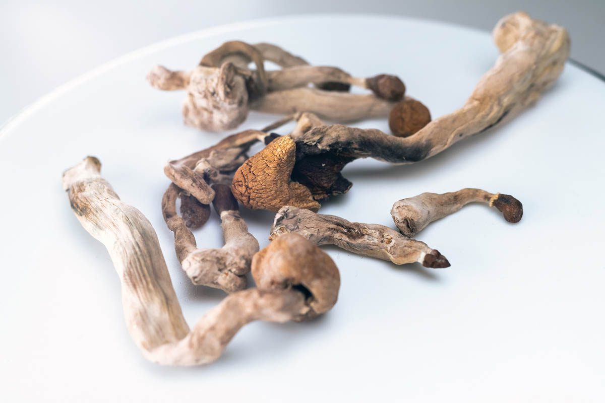
Psilocybe semilanceata gills refers to the gill structures of liberty cap mushrooms, which are critical anatomical features for accurate species identification. The gills of Psilocybe semilanceata are distinctive microscopic and macroscopic structures that help differentiate this species from toxic look-alikes and other mushroom species. This comprehensive guide covers everything you need to know about Psilocybe semilanceata gills, including structure, appearance, characteristics, identification, and comparison to other species.
What Are Mushroom Gills?
Gill Definition: Gills (or lamellae) are thin, blade-like structures located on the underside of a mushroom’s cap. These structures are the primary reproductive organs of the fungus, containing millions of microscopic spores that are released into the air for dispersal and reproduction.
Function of Gills:
- Spore Production: Gills contain basidia (spore-producing cells)
- Spore Release: Spores are released from gill surfaces
- Spore Dispersal: Wind carries spores to new locations
- Reproduction: Essential for fungal reproduction
- Identification: Gill characteristics aid species identification
Gill Anatomy:
- Hymenium: Spore-producing layer on gill surface
- Trama: Supporting tissue beneath hymenium
- Attachment Point: Where gills connect to stem
- Gill Edge: Cutting edge of gill blade
- Gill Spacing: Distance between adjacent gills
Psilocybe semilanceata Gill Characteristics
General Gill Appearance:
Color:
- Young Mushrooms: Pale brown to whitish
- Mature Mushrooms: Darker brown
- Age-Related Changes: Gills darken as mushroom matures
- Spore Maturation: Color reflects spore development stage
- Variation: Some color variation between specimens
Attachment to Stem:
- Type: Adnate to adnexed attachment
- Definition: Gills are broadly attached to the stem
- Appearance: Gills appear to be glued to stem
- Flexibility: Gills may appear slightly separated
- Consistency: Attachment relatively consistent across specimens
Gill Spacing:
- Spacing Type: Close to crowded spacing
- Distance: Gills are relatively close together
- Variation: Some variation in spacing between specimens
- Pattern: Regular, even spacing typical
- Density: High density of gills
Gill Length:
- Unequal Lengths: Gills of different lengths present
- Pattern: Longer gills alternate with shorter gills
- Terminology: Unequal or forked gills
- Identification: Helps distinguish from similar species
- Consistency: Unequal pattern typical for species
Gill Width:
- Moderate Width: Medium-width gills
- Thickness: Relatively thin gill blades
- Fragility: Gills are delicate and fragile
- Appearance: Thin, delicate appearance
- Handling: Gills easily damaged by rough handling
Gill Edge:
- Appearance: Sharp, clean cutting edge
- Condition: Typically intact and undamaged
- Fragility: Edges easily damaged
- Color: May be lighter or darker than gill face
- Erosion: Edges may appear slightly eroded in old specimens
Detailed Gill Structure
Microscopic Characteristics:
Spore-Bearing Surface (Hymenium):
- Basidia: Four-spored basidia present
- Basidioles: Sterile cells interspersed among basidia
- Spore Production: Millions of spores produced per gill
- Spore Arrangement: Spores arranged in layers
- Maturation: Spores mature progressively
Gill Tissue (Trama):
- Structure: Bilateral symmetry typical
- Composition: Hyphae arranged in parallel strands
- Thickness: Relatively thin trama
- Color: Pale, translucent appearance
- Consistency: Fragile, easily damaged
Gill Attachment:
- Connection: Gills broadly attached to stem
- Structure: Hyphae connect gill to stem tissue
- Strength: Relatively strong attachment
- Flexibility: Some flexibility in attachment
- Appearance: Gills appear glued to stem
Spore Print Characteristics
Spore Print Definition: A spore print is created by placing a mushroom cap (gill-side down) on paper or foil overnight, allowing spores to fall and create a visible print of the gill pattern.
Psilocybe semilanceata Spore Print:
Color:
- Primary Color: Dark purple-brown
- Intensity: Very dark, nearly black appearance
- Consistency: Uniform color across print
- Variation: Minimal variation in color
- Distinctive: One of most distinctive spore print colors
Pattern:
- Gill Pattern: Clear gill pattern visible
- Detail: Fine detail of gill structure reproduced
- Density: Heavy spore deposit
- Coverage: Typically complete coverage
- Clarity: Sharp, clear pattern
Quantity:
- Abundance: Abundant spore production
- Deposit: Heavy spore deposit on paper
- Visibility: Easily visible to naked eye
- Thickness: Thick, opaque deposit
- Consistency: Consistent heavy deposit
Identification Value:
- Distinctive: Dark purple-brown color highly distinctive
- Reliable: One of most reliable identification features
- Comparison: Easily compared to other species
- Confirmation: Confirms species identification
- Safety: Helps rule out toxic look-alikes
Gill Comparison to Other Species
Psilocybe cyanescens (Wavy Caps):
Similarities:
- ✓ Similar gill attachment (adnate)
- ✓ Similar gill spacing (close)
- ✓ Similar gill color progression
- ✓ Similar spore print color (dark purple-brown)
- ✓ Similar unequal gill lengths
Differences:
- ✗ Larger mushroom overall (larger gills)
- ✗ Wavy cap margin (not semilanceata)
- ✗ Slightly different gill color
- ✗ Larger spore size
- ✗ Different habitat preference
Distinguishing Features:
- Wavy caps are larger overall
- Semilanceata has bell-shaped cap
- Cyanescens has wavy cap margin
- Gill proportions differ
- Cap shape most reliable distinguisher
Mycena Species (Similar but Not Psilocybin):
Similarities:
- ✓ Similar small size
- ✓ Similar bell-shaped cap
- ✓ Similar thin stem
- ✓ Similar habitat
- ✓ Similar gill attachment
Differences:
- ✗ No blue bruising
- ✗ White spore print (not dark)
- ✗ Different gill color
- ✗ No psilocybin
- ✗ Different habitat specificity
Distinguishing Features:
- Blue bruising absent in Mycena
- Spore print color completely different
- Gill color different
- No psychoactive effects
- Habitat may differ
Galerina Species (POISONOUS – DANGEROUS):
Similarities:
- ✓ Similar small size
- ✓ Similar thin stem
- ✓ Similar habitat
- ✓ Similar cap shape
- ✓ Similar delicate appearance
Differences:
- ✗ No blue bruising (CRITICAL)
- ✗ Brown spore print (not dark purple)
- ✗ Different gill attachment
- ✗ Contains deadly amatoxins
- ✗ Highly poisonous
Distinguishing Features:
- CRITICAL: Galerina lacks blue bruising
- Spore print color completely different
- Gill attachment differs
- Galerina highly poisonous; semilanceata not toxic
- Never consume Galerina
Coprinellus Species:
Similarities:
- ✓ Similar small size
- ✓ Similar thin stem
- ✓ Similar delicate appearance
- ✓ Similar habitat
- ✓ Similar gill attachment
Differences:
- ✗ Inky gills (distinctive)
- ✗ Black spore print
- ✗ Gills dissolve with age
- ✗ No blue bruising
- ✗ No psilocybin
Distinguishing Features:
- Inky gills very distinctive
- Gills dissolve/deliquesce with age
- Black spore print completely different
- No psychoactive effects
Gill Characteristics by Maturity Stage
Young Mushrooms:
- Gill Color: Pale, whitish to light brown
- Spore Development: Spores still developing
- Gill Appearance: Gills appear lighter
- Spore Print: Lighter colored print
- Potency: Potency still developing
- Identification: Slightly more difficult
Mature Mushrooms:
- Gill Color: Medium to dark brown
- Spore Development: Spores fully mature
- Gill Appearance: Gills appear darker
- Spore Print: Dark purple-brown print
- Potency: Peak potency
- Identification: Easiest to identify
Over-Mature Mushrooms:
- Gill Color: Very dark brown to nearly black
- Spore Development: Spores fully released
- Gill Appearance: Gills appear very dark
- Spore Print: Very heavy, dark deposit
- Potency: Potency may be declining
- Identification: Easy to identify
- Condition: May show signs of age/deterioration
Gill Examination Techniques
Macroscopic Examination (Naked Eye):
Visual Inspection:
- Lighting: Use good lighting
- Angle: View from multiple angles
- Comparison: Compare to field guide images
- Documentation: Take photographs
- Notes: Record observations
Gill Characteristics to Note:
- Color and color changes
- Attachment to stem
- Spacing between gills
- Gill length variation
- Overall gill appearance
- Any damage or deterioration
Microscopic Examination:
Equipment Needed:
- Microscope: Compound microscope (400-1000x magnification)
- Slides: Microscope slides and cover slips
- Mounting Medium: Water or specialized mounting medium
- Staining: Optional staining materials
- Immersion Oil: For high magnification
Preparation:
- Sample Collection: Small piece of gill tissue
- Mounting: Place on slide with mounting medium
- Cover Slip: Apply cover slip carefully
- Pressure: Apply gentle pressure
- Examination: View under microscope
Microscopic Features:
- Basidia: Four-spored basidia visible
- Spores: Individual spore shape and size
- Hymenium: Spore-bearing layer
- Trama: Supporting tissue structure
- Cystidia: Sterile cells (if present)
Spore Print Technique:
Preparation:
- Substrate: Use white paper or aluminum foil
- Mushroom: Remove stem; place cap gill-side down
- Contact: Ensure good contact with substrate
- Cover: Cover with bowl to prevent air currents
- Time: Leave overnight (8-12 hours)
Examination:
- Removal: Carefully remove mushroom
- Pattern: Observe gill pattern in spore deposit
- Color: Note spore print color
- Density: Assess spore deposit density
- Photography: Document with photograph
Gill Characteristics in Identification Key
Identification Flowchart:
- Blue Bruising Present?
- ✓ Yes → Continue to step 2
- ✗ No → Not Psilocybe semilanceata
- Spore Print Color?
- ✓ Dark purple-brown → Continue to step 3
- ✗ White, brown, or black → Different species
- Cap Shape?
- ✓ Bell-shaped with pointed apex → Continue to step 4
- ✗ Different shape → Different species
- Gill Attachment?
- ✓ Adnate (broadly attached) → Continue to step 5
- ✗ Different attachment → Different species
- Gill Spacing?
- ✓ Close to crowded → Continue to step 6
- ✗ Distant or widely spaced → Different species
- Gill Length?
- ✓ Unequal (forked) → Likely Psilocybe semilanceata
- ✗ All equal → Different species
Confirmation:
- All characteristics match → Psilocybe semilanceata
- One or more don’t match → Different species
- Never consume without 100% certainty
Safety Considerations
Identification Importance:
- ⚠ Accurate identification critical for safety
- ⚠ Toxic look-alikes exist
- ⚠ Galerina species highly poisonous
- ⚠ Misidentification can be fatal
- ⚠ Never consume without certainty
Verification Steps:
- ✓ Use multiple field guides
- ✓ Consult experienced foragers
- ✓ Take spore print
- ✓ Examine gills carefully
- ✓ Check all characteristics
- ✓ Never consume without 100% certainty
Red Flags:
- ⚠ No blue bruising → Not semilanceata
- ⚠ Wrong spore print color → Different species
- ⚠ Different cap shape → Different species
- ⚠ Wrong gill attachment → Different species
- ⚠ Any uncertainty → Don’t consume
FAQ: Psilocybe semilanceata Gills
Q: What color are Psilocybe semilanceata gills? A: Young gills are pale brown to whitish; mature gills are medium to dark brown. Gills darken as the mushroom matures.
Q: How are Psilocybe semilanceata gills attached to the stem? A: Gills are adnate to adnexed, meaning they are broadly attached to the stem and appear glued to it.
Q: What is the spore print color of Psilocybe semilanceata? A: Dark purple-brown, nearly black. This is one of the most distinctive and reliable identification features.
Q: How do you take a spore print? A: Place the mushroom cap (gill-side down) on white paper or foil, cover with a bowl, and leave overnight. The spores will create a visible print.
Q: How do Psilocybe semilanceata gills compare to Psilocybe cyanescens? A: Very similar, but cyanescens is larger overall. The most reliable distinguisher is the cap shape: semilanceata has a bell shape; cyanescens has a wavy margin.
Q: How can you distinguish Psilocybe semilanceata from Mycena species? A: Mycena lacks blue bruising and has a white spore print. Semilanceata has blue bruising and dark purple-brown spore print.
Q: What is the most dangerous look-alike to Psilocybe semilanceata? A: Galerina species are highly poisonous and contain deadly amatoxins. The critical distinguisher is that Galerina lacks blue bruising.
Q: Are Psilocybe semilanceata gills edible? A: Psilocybe semilanceata is not toxic, but it is illegal to possess in most jurisdictions. Consumption carries legal and health risks.
Q: How do you examine gills under a microscope? A: Prepare a small piece of gill tissue on a microscope slide with mounting medium, apply a cover slip, and examine under 400-1000x magnification.
Q: What is the most reliable way to identify Psilocybe semilanceata? A: Combination of characteristics: bell-shaped cap with pointed apex, blue bruising, adnate gill attachment, close gill spacing, dark purple-brown spore print, and unequal gill lengths.
Legal Disclaimer
Important Legal Notice:
- Psilocybe semilanceata is a Class A drug in most jurisdictions
- Possession is illegal with serious criminal penalties
- This guide is for educational and identification purposes only
- It does not encourage or provide instructions for illegal activity
- Individuals are responsible for understanding and complying with local laws
- Consult legal counsel regarding drug laws
- Consequences of illegal possession can be severe
Conclusion
Psilocybe semilanceata gills are distinctive anatomical structures that serve as critical identification features for this wild psilocybin mushroom species. The gills’ characteristics—including their pale-to-dark-brown color, adnate attachment to the stem, close spacing, unequal lengths, and most importantly, the dark purple-brown spore print they produce—make them one of the most reliable ways to distinguish Psilocybe semilanceata from similar-looking species, including toxic look-alikes like Galerina.